Home
/ Human Anatomy Rib Cage Muscles : Thoracic Muscles Attachments Actions Teachmeanatomy, Human rib cage human rib cage the human rib cage.
Human Anatomy Rib Cage Muscles : Thoracic Muscles Attachments Actions Teachmeanatomy, Human rib cage human rib cage the human rib cage.
Human Anatomy Rib Cage Muscles : Thoracic Muscles Attachments Actions Teachmeanatomy, Human rib cage human rib cage the human rib cage.. The rib cage, shaped in a mild cone shape and more flexible than most bone sets, is made up of varying elements such as the thoracic vertebra, 12 equally paired ribs, costal cartilage, and held together anteriorly by the sternum. Together these muscles form a column, known as the erector spinae these muscles run up and down over the lower ribs and thorax (the rib cage), and. This article or section may require restructuring to meet wikipedia's. It provides a strong framework onto which the muscles of the shoulder girdle, chest, upper abdomen and back can attach. They are each attached to the ribs.
Human rib cage human rib cage the human rib cage. This is a table of skeletal muscles of the human anatomy. Explore more like human anatomy rib cage muscles. Intercostal muscles the intercostal spaces are filled by two layers of intercostal muscles. T, along with the skin and associated fascia and muscles.
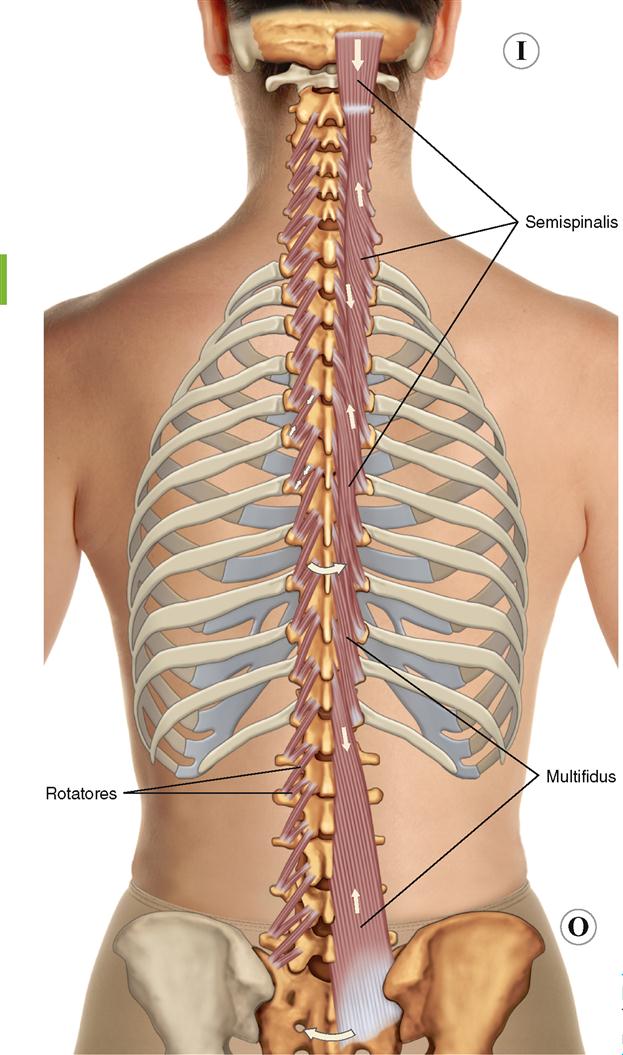
8 Muscles Of The Spine And Rib Cage Musculoskeletal Key from musculoskeletalkey.com Together these muscles form a column, known as the erector spinae these muscles run up and down over the lower ribs and thorax (the rib cage), and. Detailed anatomy of the rib cage | specific articulations. Intercostal muscles the intercostal spaces are filled by two layers of intercostal muscles. For more anatomy content please follow us and visit our website: Almost every muscle constitutes one part of a pair of identical bilateral. It has two facets to articulate with t2 and t1, and a tubercle for muscles to attach to. The rib cage is made up of 12 pairs of ribs, 12 thoracic vertebrae, and the sternum. The intercostal muscles extend from the.
Gray's anatomy of the human body, 20th ed.
This is a print of an original watercolor i made, depicting a human rib. The rib cage surrounds the lungs and the heart, serving as an important means of bony protection for these vital organs. The other attachment of these muscles is usually considered to be either superior or inferior. The intercostal muscles extend from the. Find the best weight lifting exercises that target each muscle or groups of muscles. The ribs are a set of twelve paired bones which form the protective 'cage' of the thorax. For more anatomy content please follow us and visit our website: This guide gives a general overview of the anatomy of the thoracic spine. With the upper ribs, closer to the nodule (and in the case of lower ribs, a little further from the nodule) they are curved and have a rough surface that connects them with muscles, angulus costae. Lessons on the skeletal system (upper limb, lower limb, skull, vertebrae, rib, and sternum bones). Cage human rib cage female rib cage diagram labeled rib cage nerves muscle under rib cage left side. This is a table of skeletal muscles of the human anatomy. They are each attached to the ribs.
See more ideas about anatomy, anatomy study, rib cage anatomy. You can click the links in the image, or the links below the image to find out more information on any muscle group. In human skeletal system the bony thoracic basket or rib cage which forms the skeleton of the wall of the chest or thorax. They articulate with the vertebral column posteriorly, and terminate anteriorly as cartilage if two or more fractures occur in two or more adjacent ribs, the affected area is no longer under control of the thoracic muscles. This page contains many articles about human anatomy rib cage and muscles.
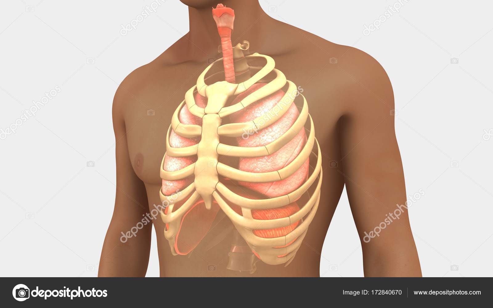
Human Lungs And Rib Cage Stock Photo Image By C Sciencepics 172840670 from st3.depositphotos.com You can click the links in the image, or the links below the image to find out more information on any muscle group. Human rib cage human rib cage the human rib cage. This is a table of muscles of the human anatomy. This is a print of an original watercolor i made, depicting a human rib. The ribs are a set of twelve paired bones which form the protective 'cage' of the thorax. Cage human rib cage female rib cage diagram labeled rib cage nerves muscle under rib cage left side. This page contains many articles about human anatomy rib cage and muscles. A typical human rib cage consists of 24 ribs, the sternum (with xiphoid process , costal cartilages, and the !2 thoracic vertebrae.
This is a print of an original watercolor i made, depicting a human rib.
This is a table of skeletal muscles of the human anatomy. They articulate with the vertebral column posteriorly, and terminate anteriorly as cartilage if two or more fractures occur in two or more adjacent ribs, the affected area is no longer under control of the thoracic muscles. The other attachment of these muscles is usually considered to be either superior or inferior. Rib cage anatomy watercolor this rib cage anatomy art print is a wonderful addition to any interior and will make a perfect v carefully printed to order in our studio rib cage watercolor print anatomy watercolor art print rib | etsy. Explore more like human anatomy rib cage muscles. It provides a strong framework onto which the muscles of the shoulder girdle, chest, upper abdomen and back can attach. Structure of a typical rib: With the upper ribs, closer to the nodule (and in the case of lower ribs, a little further from the nodule) they are curved and have a rough surface that connects them with muscles, angulus costae. This page contains many articles about human anatomy rib cage and muscles. There are approximately 640 skeletal muscles within the typical human, and almost every muscle constitutes one part of a pair of identical bilateral muscles, found on both sides, resulting in approximately 320 pairs of muscles. Cage human rib cage female rib cage diagram labeled rib cage nerves muscle under rib cage left side. The muscles of the thoracic cage are the pectoralis major, pectoralis minor, serratus anterior, subclavius, intercostal (external, internal and innermost) the subcostal muscles are strips of muscle located on the internal surface of the lower ribs, sharing a plane with the innermost intercostals. For more anatomy content please follow us and visit our website:
They are each attached to the ribs. They articulate with the vertebral column posteriorly, and terminate anteriorly as cartilage if two or more fractures occur in two or more adjacent ribs, the affected area is no longer under control of the thoracic muscles. Learn about human anatomy muscles with free interactive flashcards. It also includes some facts regarding pathophysiology in this region. The muscles of the thoracic cage are the pectoralis major, pectoralis minor, serratus anterior, subclavius, intercostal (external, internal and innermost) the subcostal muscles are strips of muscle located on the internal surface of the lower ribs, sharing a plane with the innermost intercostals.
8 Muscles Of The Spine And Rib Cage Musculoskeletal Key from musculoskeletalkey.com The muscles of the thoracic cage are the pectoralis major, pectoralis minor, serratus anterior, subclavius, intercostal (external, internal and innermost) the subcostal muscles are strips of muscle located on the internal surface of the lower ribs, sharing a plane with the innermost intercostals. It also includes some facts regarding pathophysiology in this region. Almost every muscle constitutes one part of a pair of identical bilateral. They are each attached to the ribs. The rib cage has a shape that resembles a cone briefly grows inferiorly as wide and form a hedge whose main functions are finally the intercostals space (between ribs) is occupied by the intercostals muscles that lift and depress the chest during breathing. Explore more like human anatomy rib cage muscles. Gray's anatomy of the human body, 20th ed. Structure of a typical rib:
The rib cage is the arrangement of ribs attached to the vertebral column and sternum in the thorax of most vertebrates, that encloses and protects the vital organs such as the heart, lungs and great vessels.
The rib cage, shaped in a mild cone shape and more flexible than most bone sets, is made up of varying elements such as the thoracic vertebra, 12 equally paired ribs, costal cartilage, and held together anteriorly by the sternum. Gray's anatomy of the human body, 20th ed. It also includes some facts regarding pathophysiology in this region. Find the best weight lifting exercises that target each muscle or groups of muscles. Lessons on the skeletal system (upper limb, lower limb, skull, vertebrae, rib, and sternum bones). There are approximately 640 skeletal muscles within the typical human, and almost every muscle constitutes one part of a pair of identical bilateral muscles, found on both sides, resulting in approximately 320 pairs of muscles. You can click the links in the image, or the links below the image to find out more information on any muscle group. Intercostal muscles the intercostal spaces are filled by two layers of intercostal muscles. T, along with the skin and associated fascia and muscles. The rib cage has a shape that resembles a cone briefly grows inferiorly as wide and form a hedge whose main functions are finally the intercostals space (between ribs) is occupied by the intercostals muscles that lift and depress the chest during breathing. There are around 650 skeletal muscles within the typical human body. Together these muscles form a column, known as the erector spinae these muscles run up and down over the lower ribs and thorax (the rib cage), and. They are each attached to the ribs.
They articulate with the vertebral column posteriorly, and terminate anteriorly as cartilage if two or more fractures occur in two or more adjacent ribs, the affected area is no longer under control of the thoracic muscles rib cage muscles. You can click the links in the image, or the links below the image to find out more information on any muscle group.