Home
/ Pelvis Muscles Mri Anatomy / The Pelvis Ct Anatomy Mp4 Youtube - Pelvic wall muscles include the anterior and inferior obturator internus, and the posterior iliacus, piriformis, and psoas major (figures 8 and 9).
Pelvis Muscles Mri Anatomy / The Pelvis Ct Anatomy Mp4 Youtube - Pelvic wall muscles include the anterior and inferior obturator internus, and the posterior iliacus, piriformis, and psoas major (figures 8 and 9).
Pelvis Muscles Mri Anatomy / The Pelvis Ct Anatomy Mp4 Youtube - Pelvic wall muscles include the anterior and inferior obturator internus, and the posterior iliacus, piriformis, and psoas major (figures 8 and 9).. Conclusions • the primary imaging modalities for the abdomen and pelvis are plain film, ultrasound, and ct. 48 adductor longus muscle this muscle is the most. Key facts about the muscles of the pelvic floor. A pelvis mri (magnetic resonance imaging) scan is an imaging test that uses a machine with powerful magnets and radio waves to create pictures this mri pelvis cross sectional anatomy tool is absolutely free to use. The pelvis and the pelvic floor muscles seal the abdominal and pelvic cavity in a caudal direction;
The majority of the time, these muscular tissues are quite solid and also are able to support the pelvic bone. The muscle originates from the body of the pubis and attaches to the pectineal line and proximal part of the linea aspera of femur. The pelvic diaphragm is composed of the ischiococcygeus muscle and levator ani muscle, the latter of which consists of the iliococcygeus, puborectalis, and pubococcygeus muscles. Anteriorly, pubocervical fibromuscularis is attached to the levator ani using arcus tendineus fascia pelvis (fig. The pelvis and the pelvic floor muscles seal the abdominal and pelvic cavity in a caudal direction;
Pelvis Perineum Anatomy Ppt Download from slideplayer.com The pelvic diaphragm is composed of the ischiococcygeus muscle and levator ani muscle, the latter of which consists of the iliococcygeus, puborectalis, and pubococcygeus muscles. Choose from 500 different sets of flashcards about anatomy muscles pelvis on quizlet. If these muscular tissues end up being weak. Use the mouse scroll wheel to move the images up and down alternatively use the tiny arrows (>>) on both side of the image to move the images.>>) on both side of the image to move the images. Use the mouse scroll wheel to move the images up and down alternatively use the tiny arrows (>>) on both side of the image to move the images.>>) on both side of the image to move the images. Key facts about the muscles of the pelvic floor. Case contributed by assoc prof craig hacking. A pelvis mri (magnetic resonance imaging) scan is an imaging test that uses a machine with powerful magnets and radio waves to create pictures this mri pelvis cross sectional anatomy tool is absolutely free to use.
They form a large sheet of skeletal muscle that is thicker in some areas than in others.
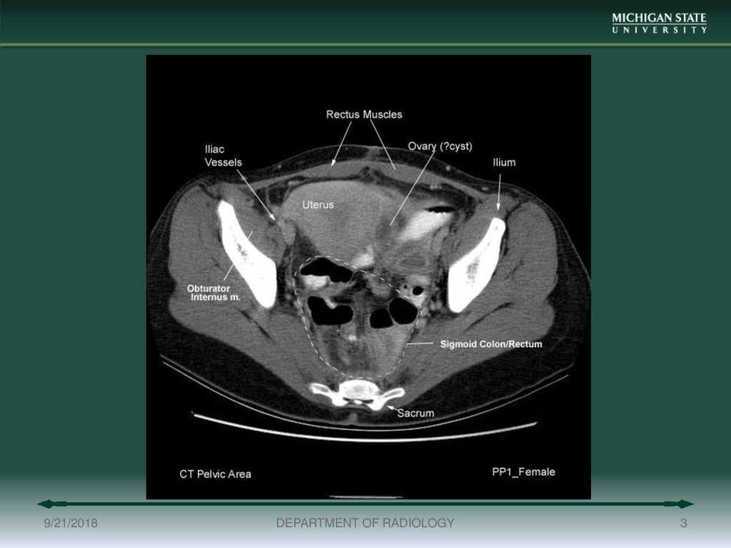
Posted by kenneth on november 18, 2019. Mri is the modality of choice in assessing the complex perineal lesions and their relations to the adjacent structures. 6.1a, b) is a bony ring consisting of paired innominate bones, the sacrum and coccyx. Anatomically, the pelvis can be divided into true and false pelvis by an oblique line that extends from the sacral promontory along the anterior aspect of s1 to the symphysis pubis. Pelvic muscle anatomy chart, pelvic muscle anatomy male, pelvic muscle anatomy pdf, pelvic muscles anatomy axial, pelvic muscular anatomy ct, human muscles, pelvic muscle anatomy chart, pelvic muscle anatomy male, pelvic muscle anatomy pdf, pelvic muscles anatomy axial, pelvic muscular anatomy ct. Magnetic resonance imaging (mri) devices can provide direct transverse, sagittal, and coronal plane images. If these muscular tissues end up being weak. Wasnik, mbbs, mda, michael b. Use the mouse scroll wheel to move the images up and down alternatively use the tiny arrows (>>) on both side of the image to move the images.>>) on both side of the image to move the images. Mri anatomy images of the abdomen. Dotted line in a ) show the anatomy of the. The innominate bones articulate with each other anteriorly and with the sacrum posteriorly. The majority of the time, these muscular tissues are quite solid and also are able to support the pelvic bone.
Mri anatomy images of the abdomen. Große auswahl an mri scans. Key facts about the muscles of the pelvic floor. The pelvis consists of an osseous ring formed by the innominate bones and sacrum, with numerous muscles for support. Posted by kenneth on november 18, 2019.
True Pelvis Pelvic Floor And Perineum Clinical Gate from i2.wp.com Mri anatomy images of the abdomen. Große auswahl an mri scans. Posted by kenneth on november 18, 2019. This mri male pelvis axial cross sectional anatomy tool is absolutely free to use. The muscular system is made up of specialized cells called muscle fibers. Mri is the modality of choice in assessing the complex perineal lesions and their relations to the adjacent structures. If these muscular tissues end up being weak. Key facts about the muscles of the pelvic floor.
The pelvic floor muscles, which are located in the pelvic outlet, make up the pelvic diaphragm, which separates the pelvic viscera from the more inferior perineal structures.
This mri male pelvis axial cross sectional anatomy tool is absolutely free to use. Use the mouse scroll wheel to move the images up and down alternatively use the tiny arrows (>>) on both side of the image to move the images.>>) on both side of the image to move the images. Anteriorly, pubocervical fibromuscularis is attached to the levator ani using arcus tendineus fascia pelvis (fig. The majority of the time, these muscular tissues are quite solid and also are able to support the pelvic bone. Anatomically, the pelvis can be divided into true and false pelvis by an oblique line that extends from the sacral promontory along the anterior aspect of s1 to the symphysis pubis. Pelvic bones are held together by the two main joints of the pelvis; This mri hip joint axial cross sectional anatomy tool is absolutely free to use. The pelvic cavity opens superiorly to, and is continuous with, the abdominal cavity through the pelvic inlet. 1 the greater, or false, pelvis is situated above this plane. E anatomy is an award winning interactive atlas of human anatomy. In this image, you will find ascending colon, appendix, psoas muscle, ureter, gluteus minimus, lumbosacral trunk, gluteus medius, gluteus maximus, sacroiliac joint, the lateral sacral crest in it. This is since pelvic muscular tissues are generally one of the major weak points in a lady's body. Key facts about the muscles of the pelvic floor.
They form a large sheet of skeletal muscle that is thicker in some areas than in others. Pelvic floor anatomy is complex and is being unraveled by means of magnetic resonance mr imaging. Right pelvic renal transplant as seen on mra. Key facts about the muscles of the pelvic floor. Although ultrasound is frequently indicated for the primary evaluation of
Ct Abdomen Pelvis Lower Axial Labeling Questions Radiology Case Radiopaedia Org from prod-images-static.radiopaedia.org Conclusions • the primary imaging modalities for the abdomen and pelvis are plain film, ultrasound, and ct. Anteriorly, pubocervical fibromuscularis is attached to the levator ani using arcus tendineus fascia pelvis (fig. The pubic symphysis and the sacroiliac joint, and reinforced by pelvic muscles. Wasnik, mbbs, mda, michael b. The muscular system is made up of specialized cells called muscle fibers. Pelvic wall muscles include the anterior and inferior obturator internus, and the posterior iliacus, piriformis, and psoas major (figures 8 and 9). Use the mouse scroll wheel to move the images up and down alternatively use the tiny arrows (>>) on both side of the image to move the images.>>) on both side of the image to move the images. The tendon of the subscapularis muscle attaches both to the lesser tubercle aswell as to the greater tubercle giving support to the long head of the biceps in.
Pelvic bones are held together by the two main joints of the pelvis;
A pelvis mri (magnetic resonance imaging) scan is an imaging test that uses a machine with powerful magnets and radio waves to create pictures of the single mri images are called slices. The pubic symphysis and the sacroiliac joint, and reinforced by pelvic muscles. The bony framework of the pelvis is called the pelvic girdle.it is composed of the two hip bones and the sacrum. E anatomy is an award winning interactive atlas of human anatomy. Conclusions • the primary imaging modalities for the abdomen and pelvis are plain film, ultrasound, and ct. This mri pelvis cross sectional anatomy tool is absolutely free to use. Pelvic bones are held together by the two main joints of the pelvis; This mri hip joint axial cross sectional anatomy tool is absolutely free to use. Mri is the modality of choice in assessing the complex perineal lesions and their relations to the adjacent structures. 12 photos of the pelvic muscle anatomy mri. They form a large sheet of skeletal muscle that is thicker in some areas than in others. Although ultrasound is frequently indicated for the primary evaluation of Anatomically, the pelvis can be divided into true and false pelvis by an oblique line that extends from the sacral promontory along the anterior aspect of s1 to the symphysis pubis.


.)